shell.hero.summary
angio.ov.summary
Control (ctrl)Inhibidor
angio.kpi.images
12
angio.kpi.wells
6
Control 3 · Inhibidor 3
angio.kpi.n_junctions
109
angio.kpi.per_image
angio.kpi.n_meshes
21,4
angio.kpi.per_image
angio.kpi.total_length
23 747
angio.kpi.per_image · µm
angio.kpi.mesh_index
281
angio.kpi.per_image · µm
Hallazgos significativos
angio.ov.findingsCondiciones
angio.ov.groups_help
Control
angio.gc.n3 · 6
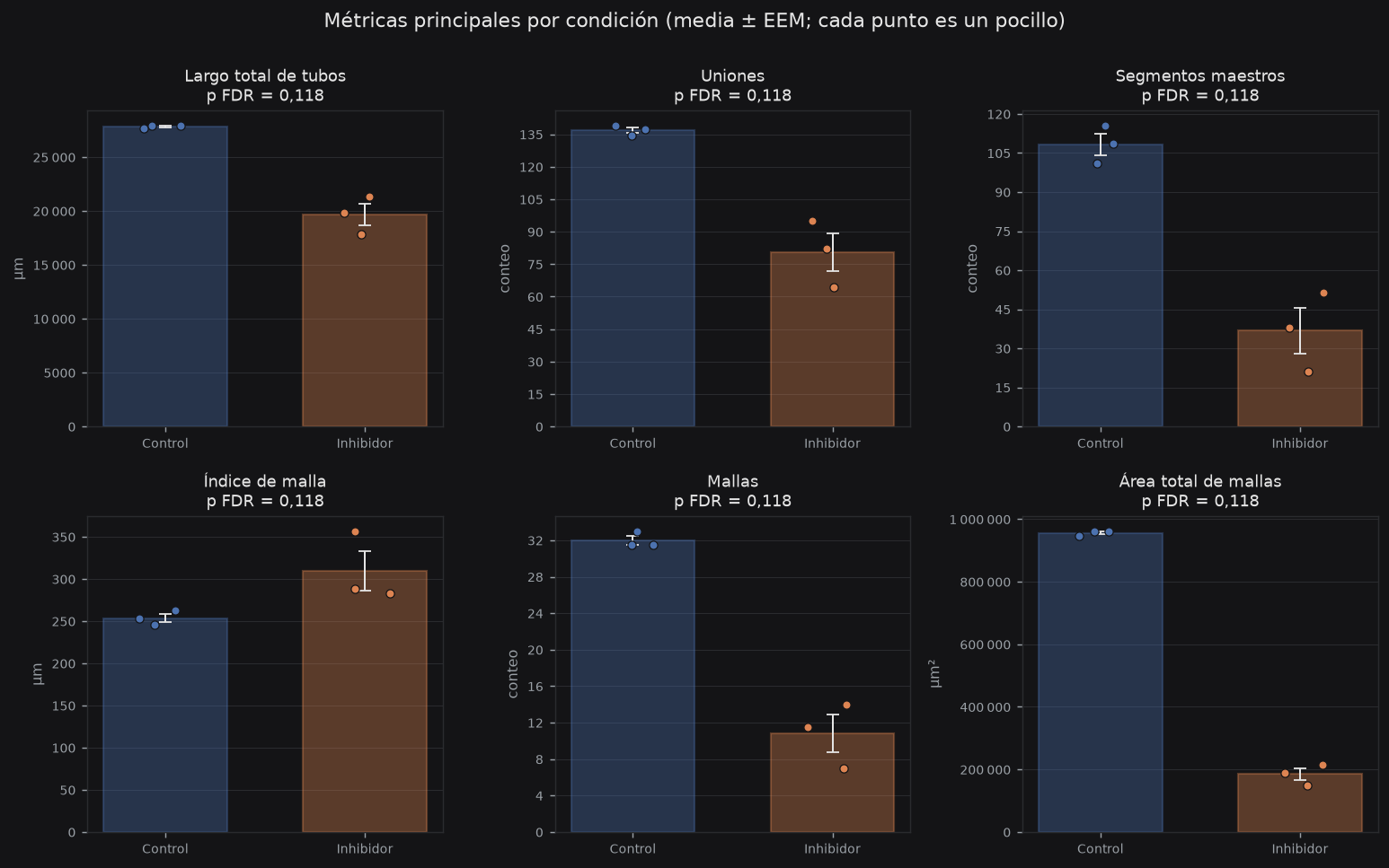
angio.m.n_junctions137 ± 2
angio.m.n_meshes32,0 ± 0,9
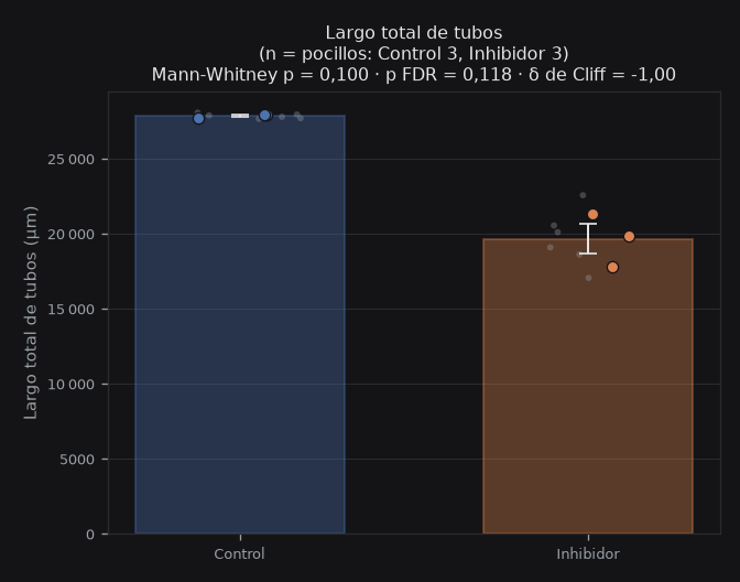
angio.m.total_length_um27 835 ± 140 µm
angio.m.total_mesh_area_um2956 465 ± 9 011 µm²
angio.m.mesh_index_um253 ± 9 µm
angio.m.n_master_segments108 ± 7
Inhibidor
angio.gc.n3 · 6
angio.m.n_junctions80,5 ± 15,3 ▼ 41 %
angio.m.n_meshes10,8 ± 3,5 ▼ 66 %
angio.m.total_length_um19 659 ± 1 755 µm ▼ 29 %
angio.m.total_mesh_area_um2184 967 ± 33 052 µm² ▼ 81 %
angio.m.mesh_index_um310 ± 41 µm ▲ 22 %
angio.m.n_master_segments36,8 ± 15,3 ▼ 66 %
Datos del experimento
angio.meta.xy
1,6
µm/px
angio.meta.units
µm · µm²
angio.meta.units_sub
angio.meta.unit
angio.meta.unit_val
angio.meta.unit_sub
angio.meta.date
—
MAPSNY · HUVEC
Método
angio.ov.method_h
angio.ov.method
Red tubular — original ↔ red reconstruida
angio.net.help
Métricas por condición
angio.mt.help
Panel de parámetros principales
Figuras por métrica
angio.mt.figs_help
angio.grp.Network
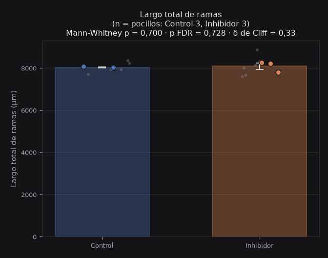
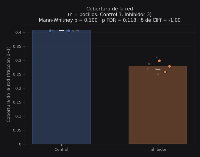
angio.grp.Topology
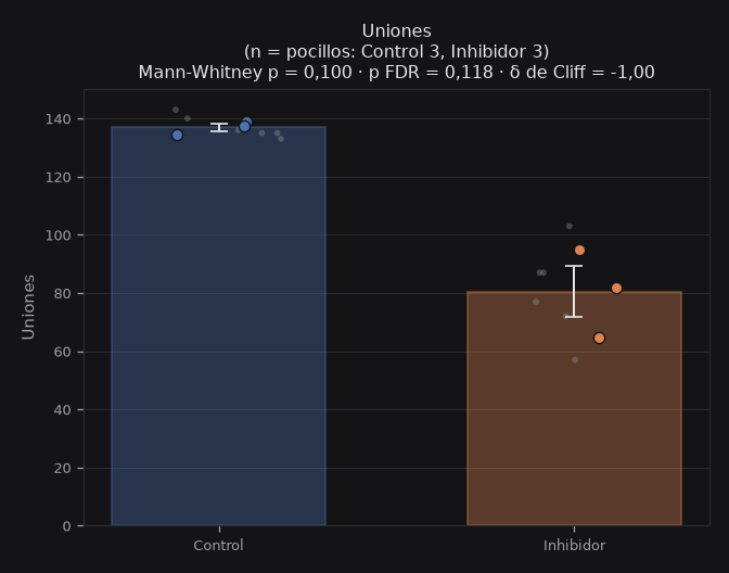
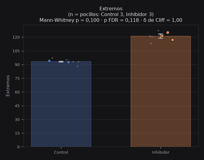
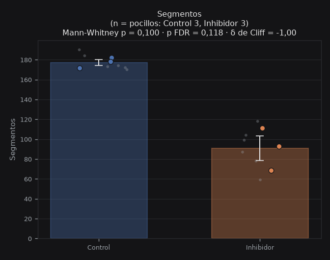
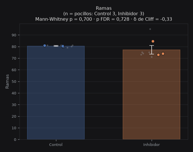
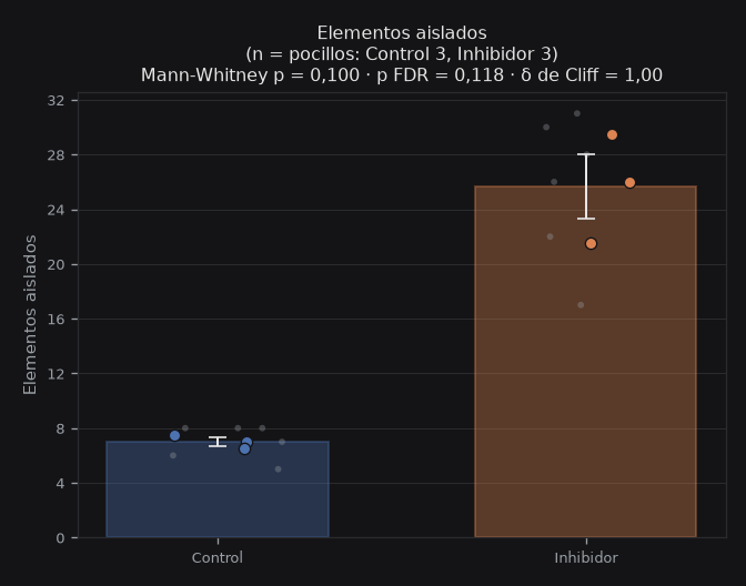
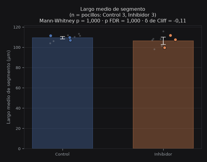
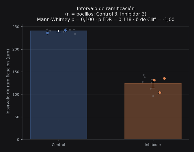
angio.grp.Master_network
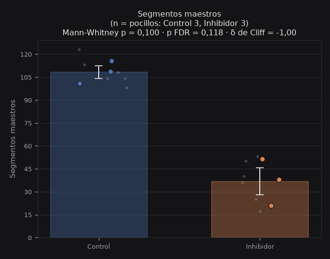
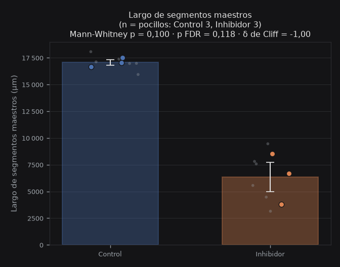
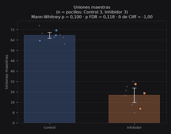
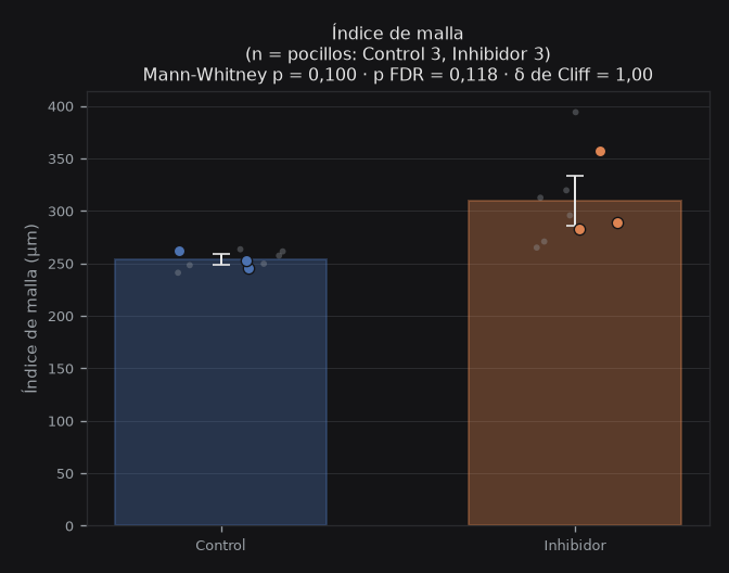
angio.grp.Meshes
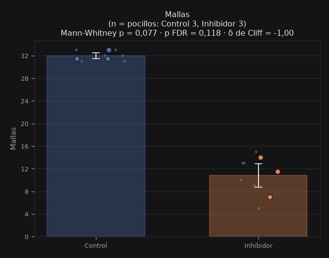
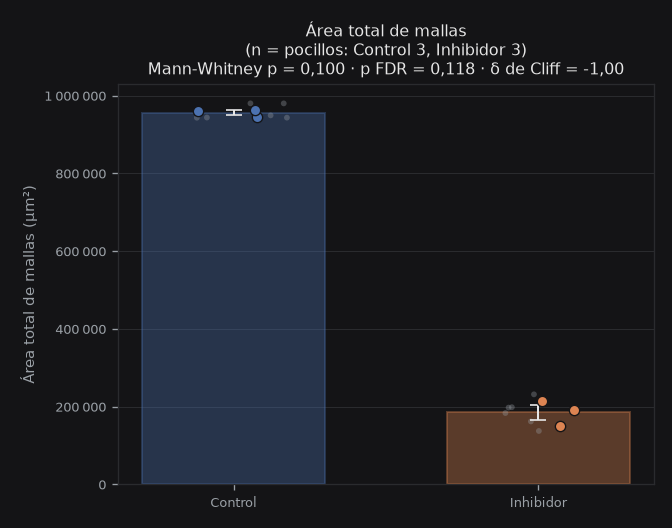
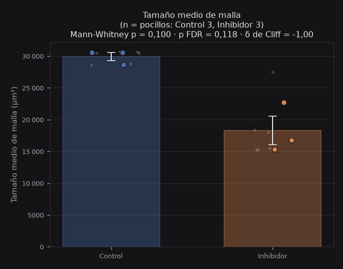
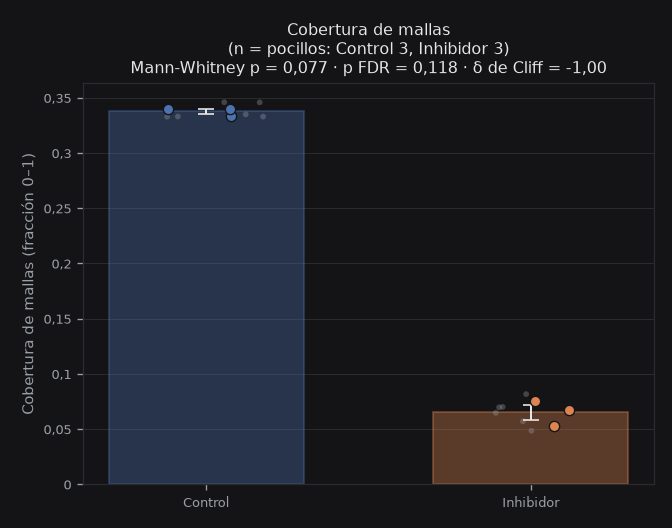
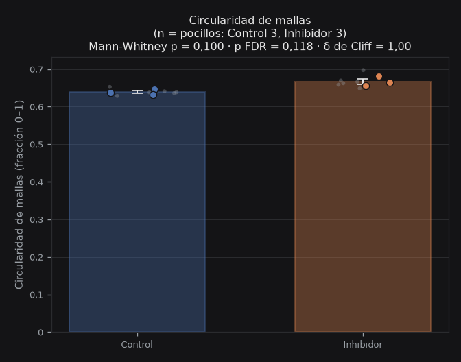
angio.grp.Density
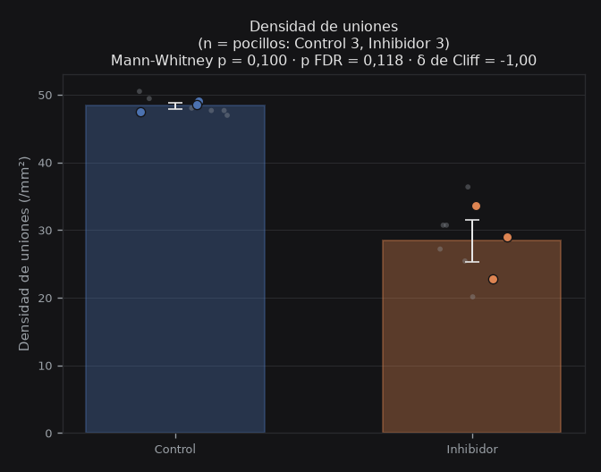
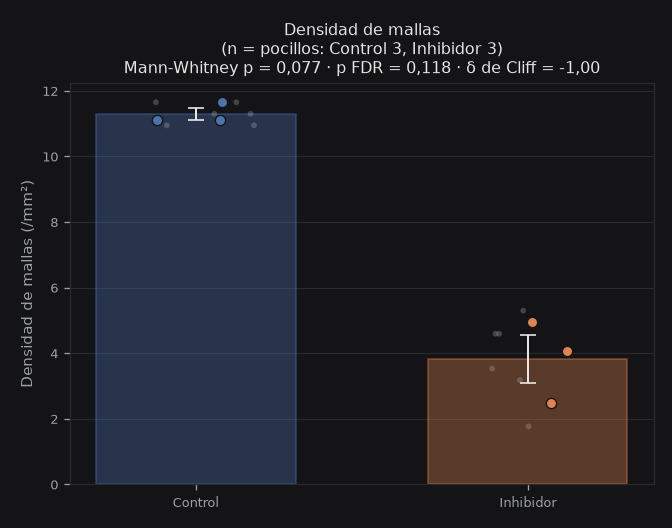
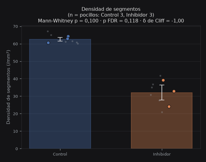
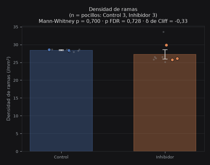
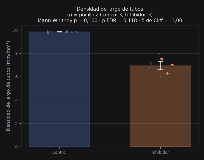
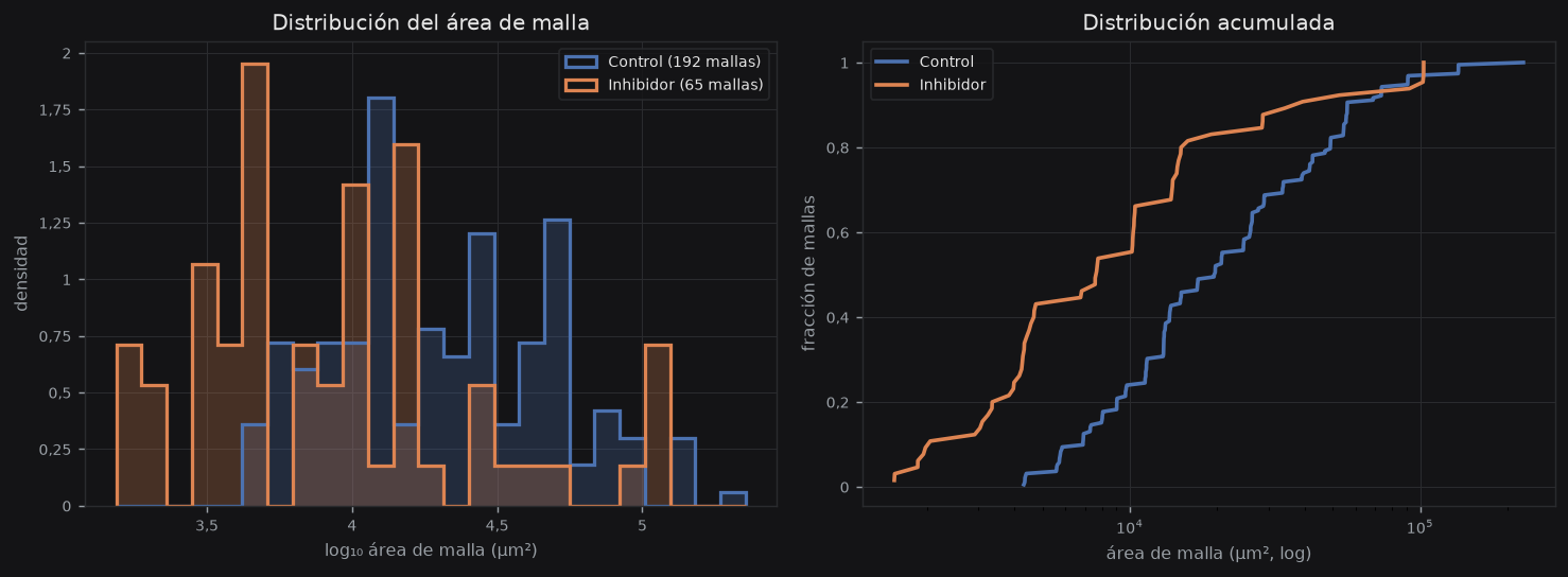
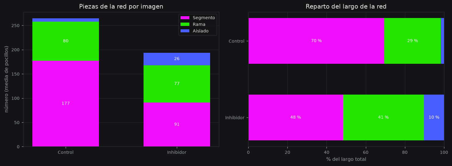
Mallas y piezas de la red
angio.mp.help
angio.mp.area_t
angio.mp.area_n
angio.mp.circ_t
angio.mp.circ_n
angio.mp.comp_t
angio.mp.comp_n
angio.mp.len_t
angio.mp.len_n
Figuras del análisis
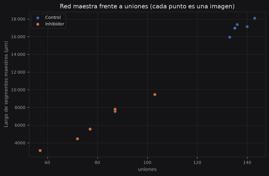
Comparación entre condiciones
angio.st.help
Informes y figuras estadísticas
Ajustes usados en este análisis
angio.set.help
| angio.set.param | angio.set.value |
|---|---|
| angio.par.grosor angio.par.grosor.h | angio.par.grosor.v |
| angio.par.sensibilidad angio.par.sensibilidad.h | angio.par.sensibilidad.v |
| angio.par.puentes angio.par.puentes.h | angio.par.puentes.v |
| angio.par.poda angio.par.poda.h | angio.par.poda.v |
| angio.par.malla_min angio.par.malla_min.h | angio.par.malla_min.v |
| angio.par.polaridad angio.par.polaridad.h | angio.par.polaridad.v |
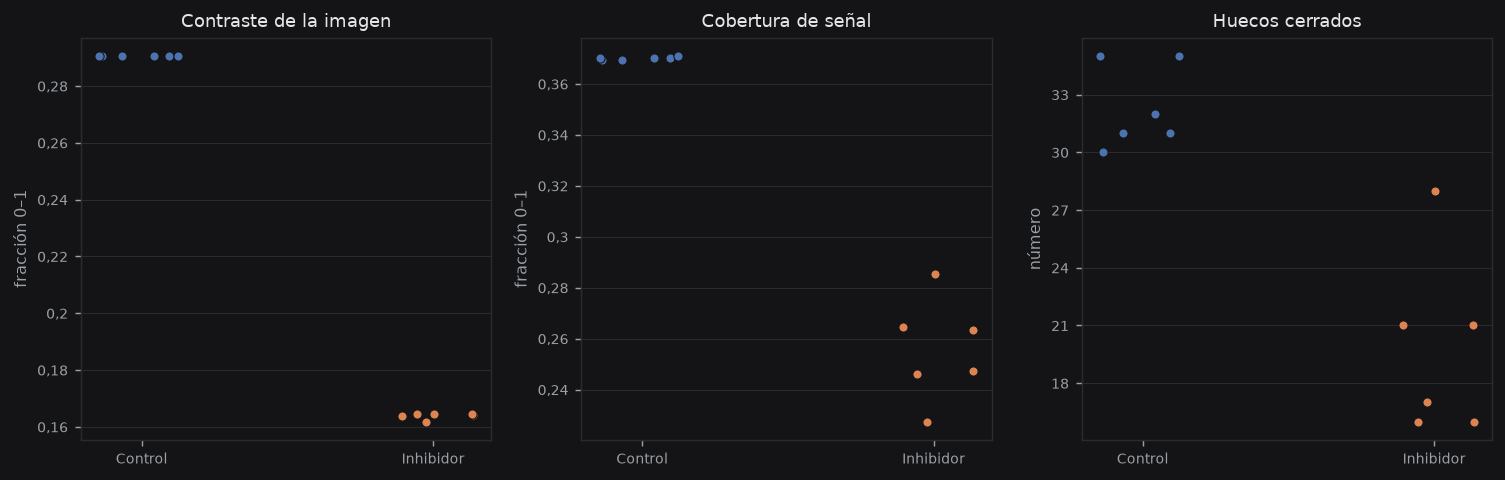
Calidad de imagen
angio.qc.k_contrast
0,23
angio.qc.median
angio.qc.k_cov
0,33
angio.qc.median
angio.qc.k_gaps
313
angio.qc.total
angio.qc.k_warn
0 / 12
angio.qc.k_warn_sub
angio.qc.help
Entrenamiento — corrige la red de una imagen
angio.tr.lead
angio.tr.help
Imágenes — una fila por imagen
angio.tb.images_help
Mallas — una fila por malla
angio.tb.meshes_help
Glossary
Statistical methodology
Referencias
- Carpentier G, Berndt S, Ferratge S, et al. (2020). Angiogenesis Analyzer for ImageJ — A comparative morphometric analysis of “Endothelial Tube Formation Assay” and “Fibrin Bead Assay”. Sci Rep 10:11568.
- Arnaoutova I, Kleinman HK (2010). In vitro angiogenesis: endothelial cell tube formation on gelled basement membrane extract. Nat Protoc 5:628–635.
- DeCicco-Skinner KL, Henry GH, Cataisson C, et al. (2014). Endothelial cell tube formation assay for the in vitro study of angiogenesis. J Vis Exp (91):e51312.
- Staton CA, Reed MWR, Brown NJ (2009). A critical analysis of current in vitro and in vivo angiogenesis assays. Int J Exp Pathol 90:195–221.
- Lazic SE (2010). The problem of pseudoreplication in neuroscientific studies. BMC Neurosci 11:5.
Downloads
shell.dl.pdf EN
SCIENTIFIC_REPORT_EN.pdf · 6 MB
shell.dl.pdf ES
SCIENTIFIC_REPORT_ES.pdf · 6 MB
angio.dl.global
GLOBAL_ANGIO_METRICS_20261007_233209.csv · 7 KB
angio.dl.fields
ANGIO_FIELDS_20261007_233209.csv · 7 KB
angio.dl.segments
ANGIO_SEGMENTS_20261007_233209.csv · 353 KB
angio.dl.meshes
ANGIO_MESHES_20261007_233209.csv · 38 KB
angio.dl.cmp
GROUP_COMPARISON.json · 33 KB
angio.dl.views
views.json · 7 KB
MAPS